


SA major hospitals benefit from new life-saving neurosurgical equipment purchased from generous donation by Wilkins Family Foundation
The Wilkins Family Foundation made a generous donation which enabled the NeuroSurgical Research Foundation (NRF) to purchase over $130,000 of lifesaving neurosurgical equipment.
SA major hospitals benefit from new life-saving neurosurgical equipment purchased from generous donation by Wilkins Family Foundation
The Wilkins Family Foundation made a generous donation which enabled the NeuroSurgical Research Foundation (NRF) to purchase over $130,000 of lifesaving neurosurgical equipment for the Royal Adelaide Hospital and Women’s & Children’s Hospital.
Several years ago, Sandy and Michaels’ daughter Kristen received a brain tumour diagnosis and has since recovered and is doing well. It was this experience that prompted the family to want to give back and help others going through similar situations.
Since that time, the Wilkins Family Foundation has donated funds to purchase a variety of different life-saving neurosurgical and research equipment throughout SA hospitals and research facilities.
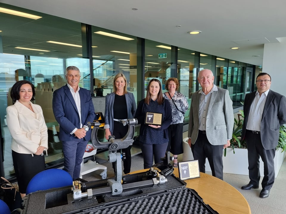
NRF President Dr Glenn McCulloch said, “We would like to sincerely thank the Wilkins Family Foundation – Michael, Sandy, Kelsey, Kristen and Mitchell for this life-saving donation. The purchase of the new neurosurgical equipment will save the lives of paediatric and adult patients in South Australia. It is a pleasure to be working together with the family to help improve patient outcomes.”
The donation by the family encouraged the equipment suppliers to make a 20% reduction in their fees. In addition, the NRF underwrote the total purchase with the top up funds required to help make the equipment purchases possible.
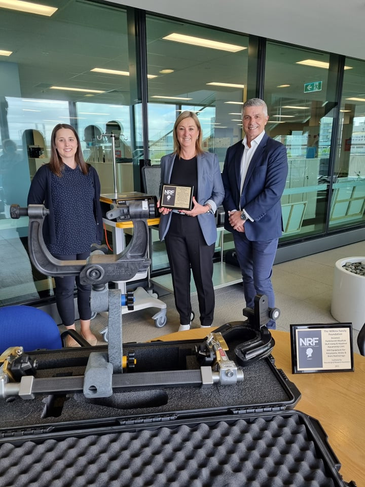
Kristen Wilkins (left), Sandy Wilkins and Michael Wilkins from the Wilkins Family Foundation at the Royal Adelaide Hospital.
The equipment purchased is outlined below:
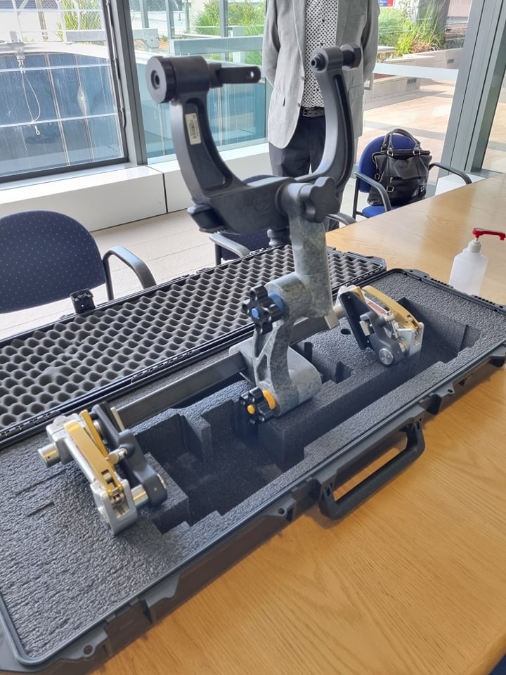
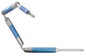
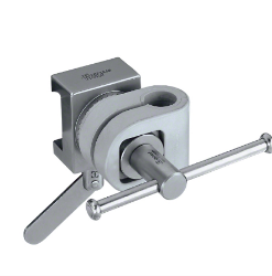
Royal Adelaide Hospital - XR2 radiolucent Mayfield skull clamp and headrest
Imaging of the brain during surgery has many advantages including ensuring completeness of removal of pathology such as brain tumour or haemorrhage before the end of surgery.
Safer and more efficient surgery ensuring the whole procedure can happen in the operating theatre without disturbing the patient and the procedure. This helps neurosurgeons achieve best outcomes for patients, particularly when surgery is performed with minimally invasive techniques.
This equipment is to be used on patients with deep brain bleeds caused by the following conditions: aneurysms, stroke, brain haemorrhage.
The older style metal skull clamp systems currently used at the RAH provide good head immobilisation for surgery but are not appropriate for clear and precise imaging during surgery as they cause artefacts making intraoperative imaging quality too poor for reliable clinical use. The XR2 radiolucent Mayfield frame is made from Carbon fibre which significantly reduces artefacts for a clearer image thus meeting modern intraoperative Imaging needs in the operating room. The radiolucent Mayfield provides rigid 3-point skeletal fixation for cranial and cervical procedures involving Digital Subtraction Angiography (DSA), Fluoroscopy, CT imaging, and MR imaging modalities.
One example of a pressing need for this equipment is the EVACUATE clinical trial: A World first clinical trial of ultra-early, minimally invasive endoscopic evacuation of brain haemorrhage led by A/Prof Amal Abou-Hamden and Prof Tim Kleinig as the lead neurosurgeon and stroke neurologist respectively at the Royal Adelaide Hospital. Intraoperative imaging during surgery allows the surgeon to ensure completeness of evacuation thus obviating the need for the patient to return to theatre for further surgery if imaging after surgery shows there is residual haemorrhage. Furthermore, the Carbon fibre Mayfield immobilisation is compatible with neuro-navigation system to allow continuous navigation during surgery without the metal interference of the current system, further improving safety and efficacy of the surgery.
Assoc/ Prof Amal Abou-Hamden
Consultant Neurosurgeon (Adult and Paediatrics)
Royal Adelaide Hospital & Women’s & Children’s Hospital
Royal Adelaide Hospital - AEX Generator for PlasmaBlade and Aquamantys
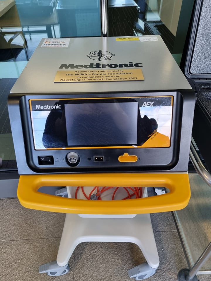
The Medtronic AEX Generator Plasmablade™ Aquamantys™ enables surgeons to cut with the precision of a scalpel and the bleeding control of traditional electrosurgery, while providing minimal thermal damage to surrounding tissue. Safer and more efficient surgery ensuring reduced surgical time, reduced total blood loss compared to traditional electrosurgery and lower transfusion rates.
Plasmablade™ dissection devices use less total energy and operate at significantly lower temperatures than traditional electrosurgery, resulting in less thermal damage to surrounding tissue.
64% lower operating temperature for cutting
45% lower operating temperature for coagulation
74% reduced thermal injury depth
Safer and more efficient surgery ensuring reduced surgical time, reduced total blood loss compared to traditional electrosurgery and lower transfusion rates. The Aquamantys™ is a bipolar sealer device providing haemostatic sealing of tissue and bone without smoke or char.
Used in Patients admitted to the RAH with aneurysms, stroke, and brain haemorrhage that develop a deep brain bleed.
Dr Stephen Santoreneos
Medical Lead of Neurosciences and Rehabilitation in CALHN
Head of unit at Women’s & Children’s Hospital
Women’s & Children’s Hospital equipment - Pneumatic Endoscopy Holder
The holder will allow an expanded use of our current neuroendoscope to allow for greater visualisation of tumours and anatomical structures during surgery in areas that are more challenging to see as they may be deeper or even just around a bend. It allows us to use the neuroendoscope as an assistance tool to our more routine open approach as the endoscope can be held in place with the holder and used as a visualization tool instead of requiring an assistant to hold it steady and in position for the duration of the surgical case. The holder also positions the endoscope in place so that we can use our hands freely to remove some tumours via the endoscope working channels itself allowing the surgical opening to be much smaller and with less morbidity.
It can be used for tumour surgery, washout of blood clots in the ventricular system in neonates, and also as an assistance in aneurysm surgery.
It will greatly improve the care we provide for our paediatric patients. This is especially since the use and application of the neuroendoscope has increased significantly. Safer and more efficient surgery ensuring the whole procedure can happen in the operating theatre without disturbing the patient and the procedure.
I am hoping that with the use of the holder, I can expand the application of neuroendoscopy in Adelaide even further to enable minimally invasive surgery especially in our paediatric population where this technology has been utilised more routinely in many centres around the world.

Dr Xenia Doorenbosch
Consultant Neurosurgeon (Adult and Paediatrics)
Royal Adelaide Hospital & Women’s & Children’s Hospital
