


Traumatic Brain injury (TBI) is the leading cause of disability and death worldwide, with as many as 60 million people suffering from a brain injury or brain damage each year.
Many of these people will develop long-lasting neuropsychiatric and cognitive impairments. And while researchers know brain injury and head injury is linked to dementia and Parkinson’s disease in later life, they still don’t know how.
Spinal Cord Injury (SCI) also leaves people disabled and needing help with everyday activities. Since there are no effective treatments, funding is urgently needed to find novel therapies to reduce this devastating impact.
With your support, we can fund these life-changing research projects.
Spinal Cord Injury (SCI) leaves patients disabled and dependent for basic daily activities. There are currently no effective treatments available for SCI and novel therapies are urgently required to reduce such devastating disability.

2023 Research Funded:
Funding: $49,660 - John Crowley Memorial Scholarship
Coagulopathy, or impaired blood clotting, complicates TBI. It worsens bleeding in the brain & confers a 9-fold increased risk of death. Current coagulopathy detection methods are slow & potentially inaccurate. ROTEM, a rapid point-of-care study, shows promise for precise & timely coagulopathy diagnosis. However, there is a lack of data to support its routine use in patients with isolated severe TBI. Our recent findings have revealed that abnormal ROTEM profile is associated with worse outcomes. We aim to investigate the underlying mechanisms & develop guidelines for utilising ROTEM in TBI management.

Mr Abhiram Devendra Hiwase, Research Assistant
University of Adelaide - Department of Health & Medical Sciences
Project: Following a concussion, the timeline of symptom recovery varies, ranging from a few weeks to those with persistent symptoms years following the initial insult. There are currently no objective measures to identify those at risk of a prolonged recovery trajectory, which is crucial to facilitate early intervention and targeted treatment. This project will utilize our clinically relevant pre-clinical model of concussion to investigate an array of promising biomarkers to determine which can predict outcome following concussion.
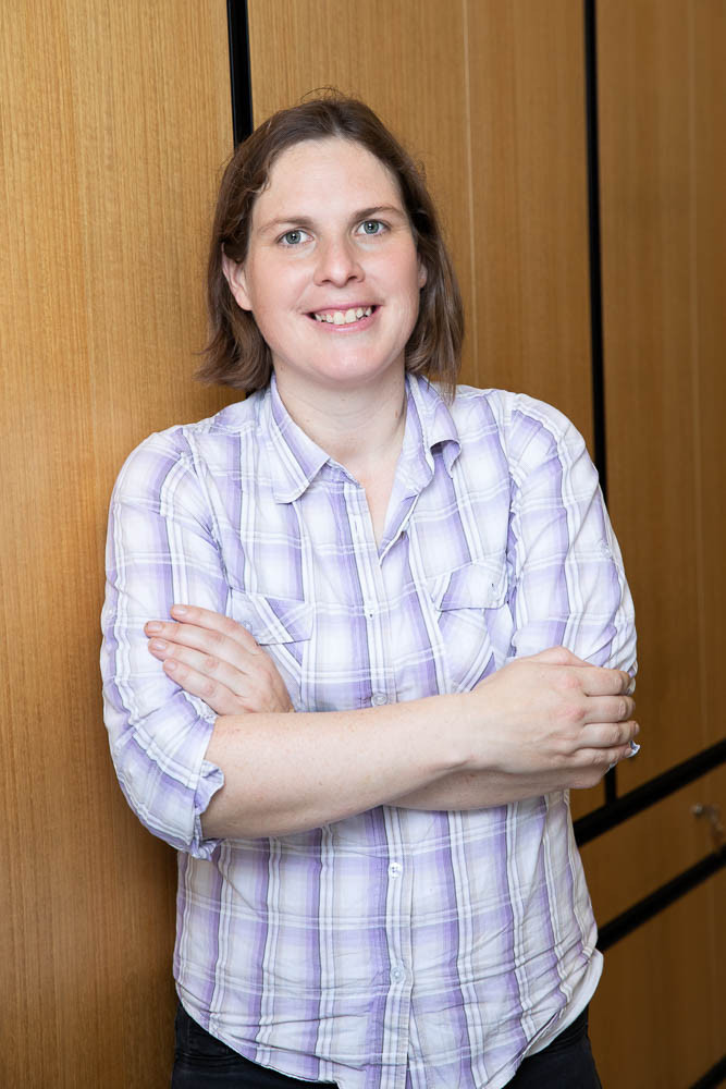
A/Prof Frances Corrigan, B Hlth Sci (Hons) PhD
Associate Lecturer, Biomedicine, University of Adelaide
2022 Research Funded:
Funding: $17,920 - John Crowley Memorial Scholarship
Chronic subdural-haematoma(cSDH) refers to a collection of old and often partially-liquefied blood clots within the skull. These collections can compresss the brain causing headache, seizures, and potentially death. They are a common neurosurgical condition in elderly. Surgical management is complicated by recurrence in up to one-third of patients. Deranged blood-clotting (coagulopathy) is suspected to contribute to this. Rotational thromboelastometry (ROTEM) is an alternative blood test to conventional coagulopathy blood tests, and may improve prediction of cSDH recurrence. We aim to perform ROTEM preoperatively for those undergoing cSDH removal, and assess whether an abnormal profile correlates with cSDH recurrence.

Mr Abhiram Devendra Hiwase, Research Assistant
University of Adelaide - Department of Health & Medical Sciences
Funding: $41,430
Project: Safe and effective control of haemorrhagic brain and vascular injury in skull base surgery is of paramount importance. Current standards of care products are limited by the depths and smaller anatomical corridors in advancing endoscopic neurosurgery.
Chitin is a naturally occurring biopolymer present in the exoskeleton of arthropods and cell wall of fungi and numerous other sources. It has antiadhesion, antimicrobial and increased clotting properties. This project aims to determine the safety and efficacy of novel beta-chitin patch in managing cerebral cortical and vascular injuries for future use in cranial skull-base surgery.

Dr Annika Mascarenhas MBBS Neurosurgery SET Trainee SET 4 (On Research Leave 2021)
University of Adelaide - Otolaryngology, Head and Neck Surgery/Neurosurgery
Funding: $15,220
Project: This study will be conducted in two phases. The primary purpose is for the development of an animal model of endoscopic small vessel handling, brain manipulation and haemorrhage control in the brain and dural closure techniques using sheep.
Once feasibility has been established with the pilot study in phase 1, then phase 2 will be the subsequent establishment of an endoscopic animal training model for ENT surgeons and Neurosurgeons for further development of operative skill.
Dr Annika Mascarenhas MBBS Neurosurgery SET Trainee SET 4 (On Research Leave 2021)
University of Adelaide - Otolaryngology, Head and Neck Surgery/Neurosurgery
Funding: $20,000
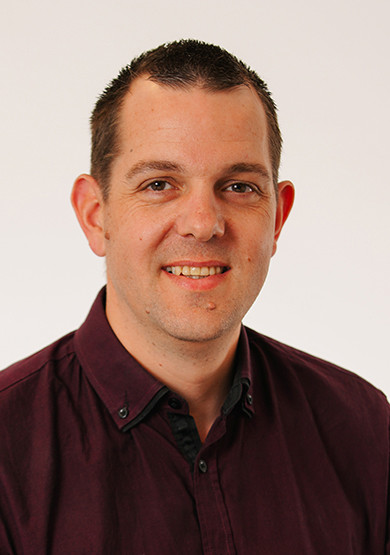
Project: Whilst hundreds of patients with potential brain injury present to the RAH every year, this study focuses on a subgroup at higher risk of neurological deterioration.
This group consists of patients who are assessed as having a moderate or severe TBI and are thus more likely to benefit from ROTEM use. According to past patient numbers, we estimate this group to be around 180 patients per year. In conjunction with our trauma colleagues, we aim to implement ROTEM testing in all patients presenting to our emergency department over the next three years with a moderate or severe TBI.
This project will enable us to investigate the relationship between TBI, ROTEM results and patient outcomes. By collecting ROTEM results, demographic data of patients and clinical outcomes, we aim to demonstrate the predictive value of ROTEM testing in this population.

Dr Adam Wells - Neurosurgeon
Royal Adelaide Hospital - Neurosurgery
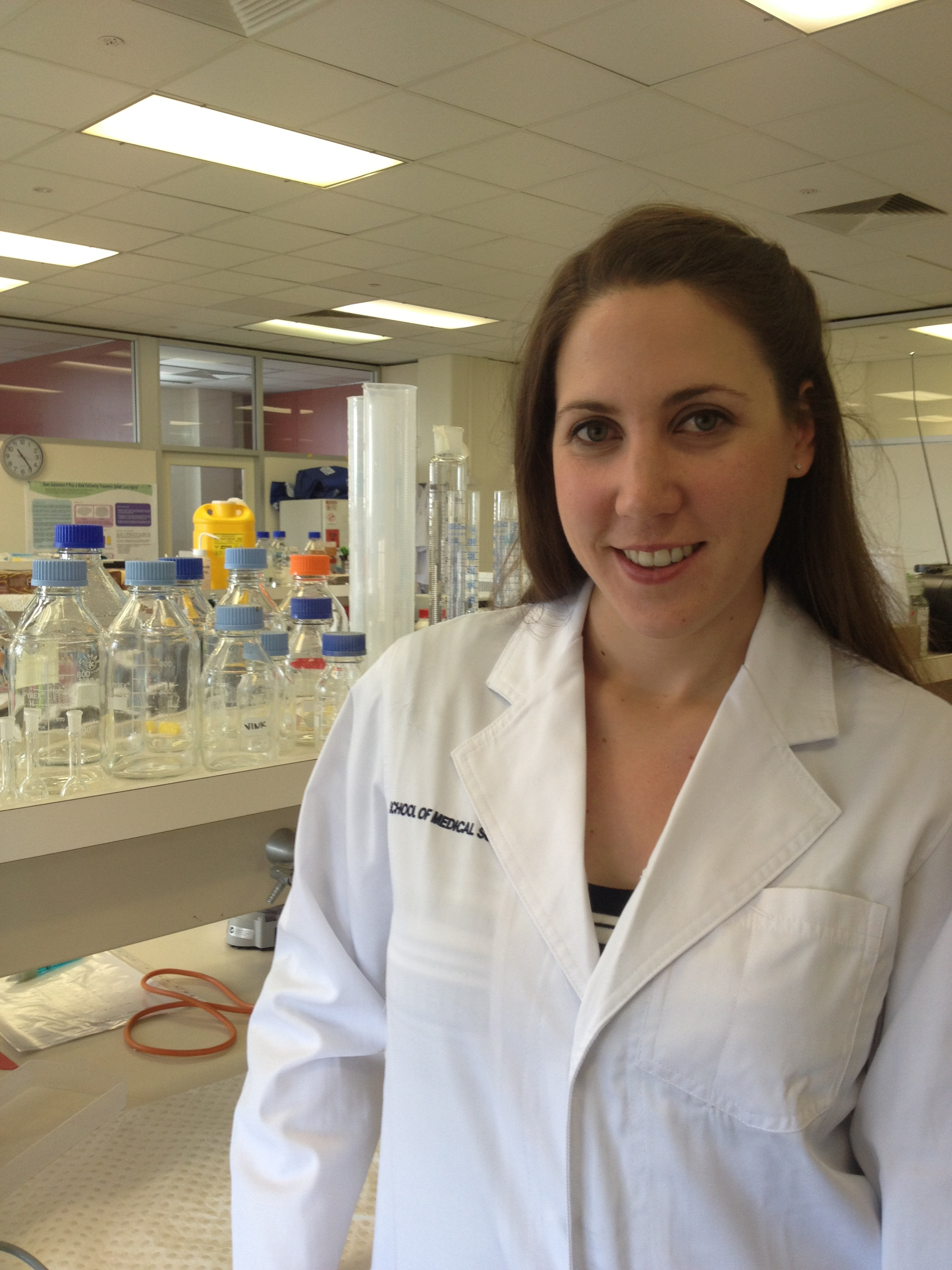
I’m a neuroscientist with a key research interest in how diet and nutrition impacts brain function at both molecular and behavioural levels. A major part of my research program involves understanding how the network of molecules that surround neurons – called the extracellular matrix – can influence cognitive function – in particular memory formation.
A specialised form of extracellular matrix wraps around neurons, which is called a ‘perineuronal net’. This structure provides both a way of stabilising the physical connections that neurons form when encoding memories, and also provides a protective shield against damage in neurodegenerative conditions – including traumatic brain injuries.
My recent research shows that poor nutrition can damage perineuronal nets and cause memory impairments. I will be investigating how the combination of brain injury and nutritional status functionally affects neurons, perineuronal nets and memory abilities using a translational rodent model.

Dr Amy Reichelt
Senior Lecturer, University of Adelaide Medical School
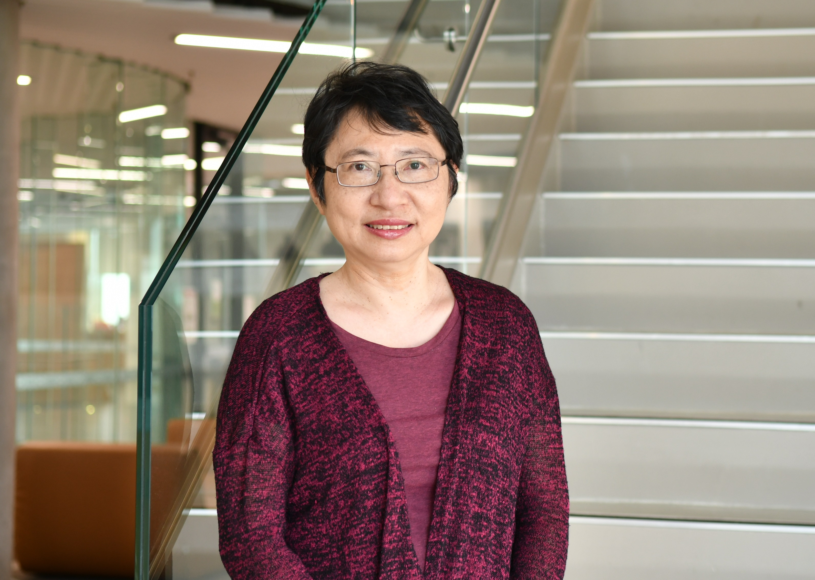
Traumatic brain injury (TBI) is a major cause of disability and mortality worldwide, with increasing recognition that it can have life-long consequences. TBI may increase the risk of dementia, with suggestions that a dose-dependent relationship might exist. TBI is associated with neuropathological changes characteristic of Alzheimer’s disease (AD). AD is clinically defined by a progressive loss of cognitive function and is the most common neurodegenerative disease and the leading cause of dementia, accounting for 75% of cases. This project will test a novel hypothesis regarding the pathogenesis of AD following TBI using immunohistochemistry and will highlight potential imaging biomarkers.
We will also establish a novel MALDI imaging technique to identify spatial perturbations in neurotransmitters caused by TBI. The neurotransmitter imaging method will be made available to the wider neuroscience research community. We will be the first to establish this cutting-edge technology in Australia working closely with its inventor.
Dr Paul J. Trim
SA Health & Medical Research Institute (SAHMRI)
Traumatic brain injury (TBI) encompasses pathophysiological changes known as secondary injury processes that often lead to worsened prognosis and outcome. To date there is no reliable methodology to predict which patients may deteriorate acutely following TBI. This project aims to investigate the immediate inflammatory response to TBI, via cerebral microdialysis, which allows direct sampling of the fluid within the brain to determine if alterations in the expression of any proteins within the inflammatory response predict the later development of brain swelling. The ability to identify these patients earlier, allows therapeutic intervention to begin which may prevent these deleterious outcomes.

Ms Lola Kaukas PhD Student
University of Adelaide
Traumatic brain injury (TBI) is the leading cause of death in individuals under the age of 45 years and survivors are often left with long-term disability. In particular, patients with post-TBI gastrointestinal dysfunction have increased morbidity and longer periods of hospitalization. Therefore, treatment modalities targeting prevention of gastrointestinal dysfunction have important clinical implications. In the current study we will characterise both the time course and nature of gastrointestinal disturbances following trauma. As such, this study will evaluate the extent of gastrointestinal disturbances, including gut injury, increased permeability and alterations in inflammatory mediators, that occur following moderate traumatic brain injury. This may lead to the identification of novel therapeutic targets to reduce gastrointestinal complications and improve TBI patient quality of life.
Assoc Prof Stuart Brierley
Mathew flinders Fellow in Gastrointestinal Neuroscience Flinders University
Mild traumatic brain injury (mTBI) is one of the most common forms of acquired brain injury, affecting millions of people around the world every year. Although once considered a short-lived injury, the potential long-term side effects of mTBI are now being increasingly recognised. Despite this, the physiological mechanisms contributing to these deficits are largely unknown, placing considerable limitations on how mTBIs are handled clinically. Using advanced neuroimaging techniques, my work aims to better understand how mTBI changes the brain, and how these changes result in ongoing functional deficits. This will allow us to develop markers of injury that can be used to track recovery from mTBI, and may eventually facilitate the design of interventions to reduce the burden of ongoing symptoms.

Dr George Opie PhD
NHMRC Early Career Fellow, Discipline of Physiology, The University of Adelaide
Parkinson’s disease (PD) is the second most common neurodegenerative disease after Alzheimer’s disease, affecting 10 million people worldwide and 1 in every 350 Australians. While the exact causes of PD are currently unknown, one risk factor is traumatic brain injury. Despite growing awareness of the link between TBI and PD, however, brain mechanisms that account for this relationship are unknown. One potential mechanism may be neuroinflammation. A potent inducer of neuroinflammation is activation of Toll-like receptor 4 (TLR4), a pattern recognition receptor broadly expressed in the central nervous system. The current study will investigate whether the development of neuroinflammation and PD-like pathology following TBI is mediated by TLR4 activation. This has the potential to shed light on the mechanism by which a major risk factor for PD may lead to disease, and may help to identify novel therapeutic targets.
While the acute effects of traumatic brain injury (TBI) are well-known, a number of individuals affected by TBI also develop chronic problems such as depression and cognitive impairment. Although the brain mechanisms of these impairments are currently unclear, persistent inflammation in the brain may play a key role.
Our current NRF-funded research projects investigate whether reducing brain inflammation immediately after injury can improve long-term outcomes in an experimental model of TBI. This work may have important consequences for the prevention of neurodegenerative diseases, such as dementia.
Assoc Prof Lyndsey Collins-Praino
Lecturer in Anatomy & Pathology, Adelaide Medical School, The University of Adelaide
Assoc Prof Lindsey Collins-Praino speaks further about her research.

2023 Research Funded:
Funding: $59,020
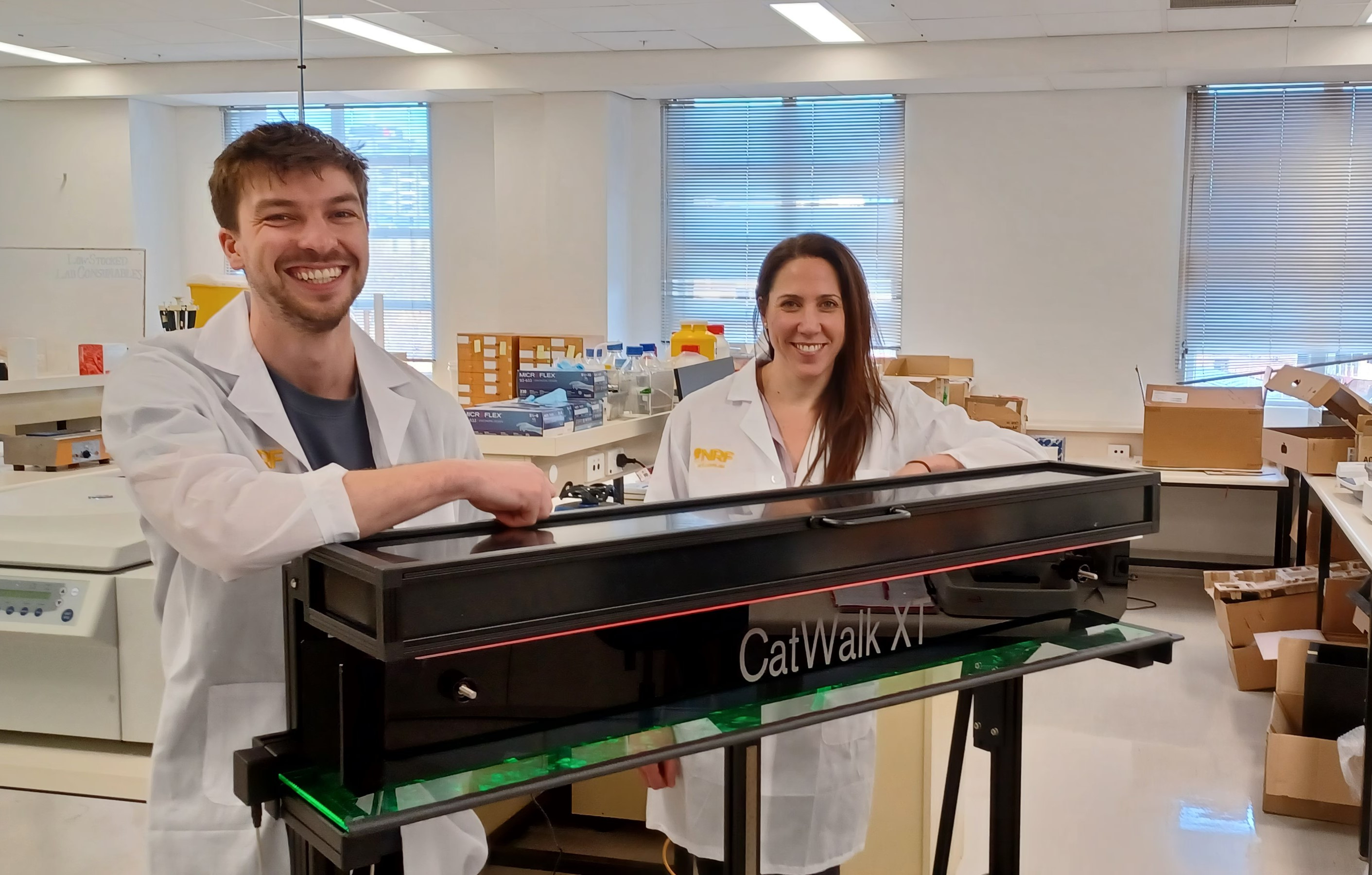
Neurological diseases or injuries can drastically impair motor function; however, these deficits can be difficult to identify and characterise in preclinical models. The Catwalk is a state-of-the-art system that will allow us to automatically quantify deficits in key motor outcomes, including walking speed, stride pattern, pressure, and coordination. This will enhance our understanding of various pathologies such as Spinal Cord Injury, Traumatic Brain Injury and Stroke, and enable us to examine potential interventions that may one day improve outcomes for people living with these conditions.
Dr Anna Leonard B.HlthSc (Hons), PhD Senior Lecturer in Anatomy & Pathology
University of Adelaide - Adelaide Medical School
Funding: $50,000
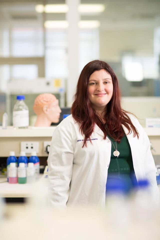
Over 15,000 Australians currently live with permanent disability following spinal cord injury (SCI). Many perceive SCI to predominantly affect an individual’s physical ability, however chronic pain develops in 75% of SCI survivors, significantly reducing quality of life. Although normal sensation is lost, the brain creates the perception of pain due to disrupted neuronal signaling. The underlying mechanisms are not well understood, and no efficacious treatments exist. Here we will investigate how a head injury, which occurs in 60% of SCI cases, facilitates development of chronic pain and evaluate the potential mechanisms at play to identify future therapeutic targets.

Dr Anna Leonard B.HlthSc (Hons), PhD Senior Lecturer in Anatomy & Pathology
University of Adelaide - Adelaide Medical School
Funding: $31,688
Project: To improve outcomes and quality of life for individuals with SCI, devices that are minimally invasive, comfortable and effectively alter pain are desperately needed. The current study will investigate a new wireless device called a 'graft antenna', which can be implanted on uninjured peripheral nerves, such as the sciatic nerve and stimulated via electromagnetic induction. We propose that weekly peripheral stimulation via this novel method will reduce neuroinflammation whilst promoting tissue repair and regeneration within the spinal cord itself.
Dr Anna Leonard B.HlthSc (Hons), PhD Senior Lecturer in Anatomy & Pathology
University of Adelaide - Adelaide Medical School
Funding: $17,328
Project: Spinal cord injury (SCI) is traumatic event that often results in profound neurological deficits, significantly impacting the individual’s physical, psychological, and social wellbeing. Up to 810 SCIs occur in Australia annually, causing a significant financial burden on the individual, family and healthcare system.
The lifetime cost per individual is estimated to be $5-9.5m, whilst the annual cost within Australia alone is >$2bn. Accordingly, SCI research is urgently needed and our research team at is expanding to meet this demand. New behavioural equipment will increase the capacity of our research, expanding into the field of chronic outcomes such as neuropathic pain.
Dr Anna Leonard B.HlthSc (Hons), PhD Senior Lecturer in Anatomy & Pathology
University of Adelaide - Adelaide Medical School
Computed tomography (CT) and magnetic resonance imaging (MRI) images are routinely used in neurosurgery for clinical decision making, pre-operative planning and post-operative follow-up.
They provide complementary information but are acquired on different imaging devices and at a separate time. This interrupts the clinical workflow, adds to health-care costs, and poses challenges in registering the images for analysis. Using innovative technologies, our aim is to synthesise spinal CT images from MRI images - providing the necessary MRI information without performing any real CT scans.
This will improve clinical workflow, reduce costs to patients and the health system, as well as help minimise patient exposure to ionising radiation due to a CT scan. The impact also extends beyond spine imaging, notably to paediatric neuroimaging, where childhood exposure to ionising radiation has demonstrable causal effects on the development of cancer in later life.
Dr Gobert Lee
Lecturer in Statistical Science
College of Science and Engineering
Flinders University
Spinal cord injury often servers the communication between the peripheral body and the brain. While the spinal cord itself has been damaged the peripheral nerves remain intact. Electrical stimulation of these nerves has been shown to promote the growth of healthy nervous tissue into and across the injury site of the spinal cord. Whilst electrical stimulation devices have shown some success in motor recovery for individuals with SCI, they remain invasive, bulky and only modestly effective for pain. To improve outcomes and quality of life for individuals with SCI, devices that are minimally invasive, comfortable and effectively alter pain are desperately needed.
The current study will investigate a new wireless device called a “graft-antenna”, which can be implanted on uninjured peripheral nerves, such as the sciatic nerve, and stimulated via electromagnetic induction. We propose that weekly peripheral stimulation via this novel method will reduce neuroinflammation whilst promoting tissue repair and regeneration within the spinal cord itself.
Dr Anna Leonard
Lecturer in Anatomy & Pathology, Adelaide Medical School, The University of Adelaide
Click here to donate to the Traumatic Brain Injury Research Appeal

